Maxillofacial Trauma: The Role of Multidetector Row Computed Tomography
Maxillofacial Trauma: The Role of Multidetector Row Computed Tomography
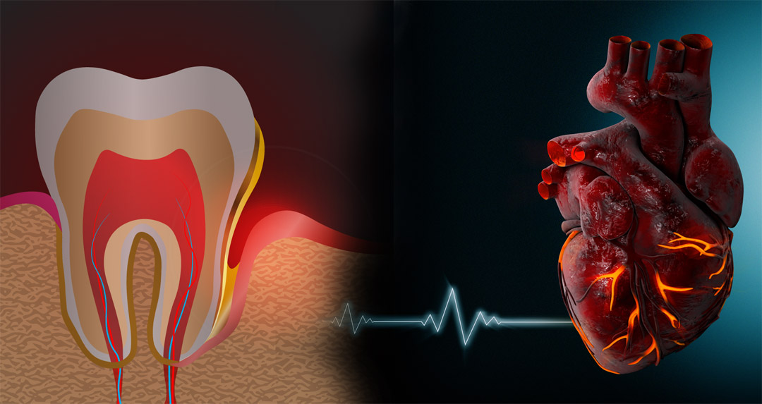
Multidetector-row computed tomography (MDCT) is a recent advancement in CT technology. Using multiple detector rows allows the imaging of a larger area. MDCT can visualize proximal and distal vessel segments. Because of its increased resolution, MDCT has the potential to improve patient care. However, it also comes with several pitfalls. This article will review the basic principles of MDCT scanners and the benefits and limitations of this advanced technology.

The advantages of multidetector row CT over conventional radiography include better soft-tissue characterization. Unlike single-detector helical CT, these images are more accurate and can help physicians distinguish between benign and malignant lesions. Its high-resolution imaging can also help in the diagnosis of inflammatory lesions and tumors in the head, neck, abdomen, and cranial regions.
Although this imaging method is not always as accurate as conventional X-rays, it is still the first choice of physicians after traumatic facial trauma. This test helps identify fractures and their complications. A multidetector computed tomography examination is highly sensitive and is an excellent choice for detecting small fractures. It is also helpful in assessing the extent of displacement and fracture sites, which is vital for accurate surgical planning.
Multidetector-row CT is an excellent tool for assessing the severity of a nasal bone fracture. Moreover, it is a valuable diagnostic tool for treating maxillofacial fractures. Despite the limitations of 3D images, a clinically useful and inexpensive 3-D CT can be used to detect even the smallest complication. This makes it more accessible than conventional X-rays.
The main advantage of this imaging technique is its sensitivity. It has better accuracy than conventional X-rays. Furthermore, it does not depend on the location of the perforation. It is also sensitive to free air. In addition, it does not rely on the presence of soft tissues. Hence, MDCT is an excellent option for diagnosing traumatic fractures in this region.
Compared to intravascular ultrasonography, multidetector-row CT is an excellent choice for identifying fractures. Its sensitivity for detecting stenoses is greater in the distal part of the body than the proximal segment. This CT technology has the potential to improve diagnostic accuracy in the face. The ability to see all artery segments in a 3D image is an important feature for patients with mid-facial fracture.
In contrast to the limited spatial resolution of CT, MDCT offers superior delineation of osseous structures. The spatial resolution of MDCT is remarkably high and allows for thin-section acquisitions. Because of the high fidelity of images, multidetector-row CT is a superior choice for diagnosis of maxillofacial fractures. But it is not without its shortcomings.
In addition to providing three-dimensional images, multidetector CT also offers three-dimensional information. It allows doctors to evaluate the aortic valve apparatus, which is crucial to a high-result in the surgery. The calcifications in the prosthesis can also affect the success of the operation. In cases of moderate aortic regurgitation, this imaging technology was found to be effective in predicting the success of the surgery.
In recent years, MDCT has become the gold-standard imaging modality for the evaluation of head and neck trauma. Its superior soft-tissue contrast and a high-resolution image of the skull base allow it to be used in certain cases. For patients with cranial nerve deficits, MDCT is also useful for assessing the extent of cranial bone fractures.
A high-resolution CT scan is crucial for accurate angiography. It allows physicians to see the whole anatomy of the patient. This is important, because the more angiographic data, the better. In addition, it can be used to diagnose plaques. It also helps physicians detect stenoses in coronary arteries. The technique can accurately determine the extent of a patient’s artery disease.
Due to the enormous socioeconomic impact of coronary artery disease, cardiac multidetector-row CT has become the standard of reference for determining the severity of a patient’s CAD. Besides the high resolution, the technology has also improved angiography’s z-axis resolution. It can be used to diagnose bowel perforation. There are two types of FD: FB and angiography.